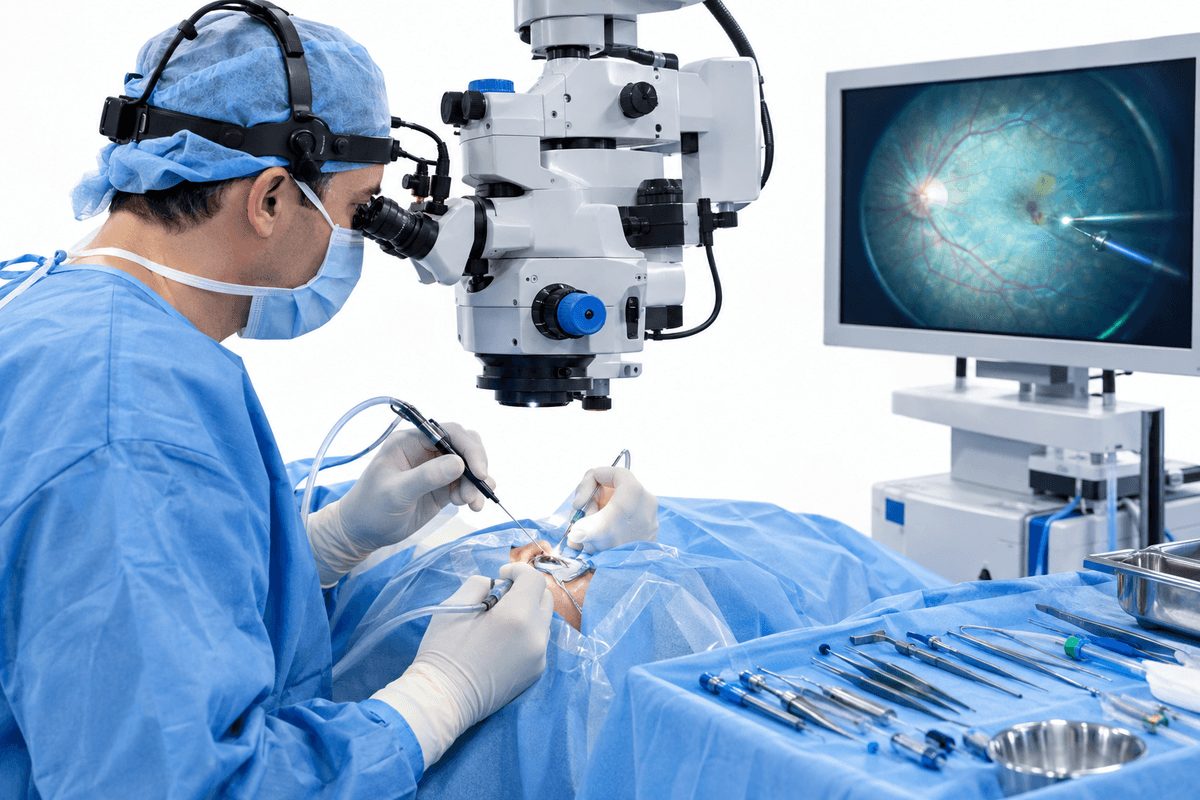
Performed by our specialist team at Shreedeep Hospital
Retinal surgery in Ahmednagar addresses serious retinal conditions including retinal detachment, macular holes, diabetic retinopathy, and epiretinal membrane. This microsurgical procedure aims to restore or preserve vision by repairing damaged retinal tissue and preventing further vision loss. Our specialist team at Shreedeep Hospital performs advanced retinal surgeries using modern vitreoretinal techniques.
We believe in honest, upfront disclosure so you can make an informed decision.
A step-by-step look at what happens during this procedure.
The vitreous gel is carefully removed from the eye using microsurgical instruments to access the retina and clear any blood or debris obstructing vision.
Damaged retinal tissue is repaired using laser photocoagulation, cryotherapy, or surgical instruments to seal tears, flatten detachments, or remove scar tissue.
A gas bubble or silicone oil may be injected into the eye to hold the retina in position against the eye wall during healing.
In cases of retinal detachment, a silicone band may be placed around the eye to indent the wall and reduce tension on the retina.
Surgical incisions are closed, antibiotic drops are applied, and the eye is patched or shielded to protect it during initial healing.
A structured, patient-first approach from first consultation to full recovery.
Shreedeep Hospital conducts detailed retinal imaging including OCT scans and fundus photography to accurately map the extent of retinal damage and plan the surgical approach.
Our specialist team utilizes modern vitreoretinal surgical equipment and techniques including micro-incision vitrectomy systems for minimal trauma and faster recovery.
Our team provides detailed positioning instructions, medication schedules, and regular follow-up examinations to monitor retinal healing and detect any complications early.
Shreedeep Hospital ensures ongoing monitoring of retinal health and manages underlying conditions like diabetes or hypertension that may affect surgical outcomes.
What to expect at each stage of your recovery journey.
The first week requires strict head positioning if gas tamponade was used, avoiding air travel, and using prescribed eye drops. Vision will be significantly blurred, and activities are restricted.
Weeks 2-6 involve gradual visual improvement as the retina heals and gas bubble absorbs. Patients can resume light activities but must avoid heavy lifting, bending, and strenuous exercise.
After 6 weeks to 6 months, most patients experience their final visual outcome. Regular monitoring continues to ensure retinal stability and address any residual issues like cataract development.
Most patients experience stabilization or improvement in vision following primary retinal detachment repair, with outcomes depending on the duration and severity of detachment.
Surgical intervention successfully reattaches the retina in the majority of cases, preventing permanent vision loss and restoring the normal anatomical structure of the eye.
Early surgical intervention halts the progression of retinal disease, preventing complications like proliferative vitreoretinopathy and complete vision loss.
Restored vision enables patients to resume daily activities, maintain independence, and significantly improves overall quality of life and functional outcomes.
Let's clear up some of the most common myths about retinal surgery.
Get a personalised assessment from an experienced specialist. Understand your options, ask your questions, and decide with confidence.